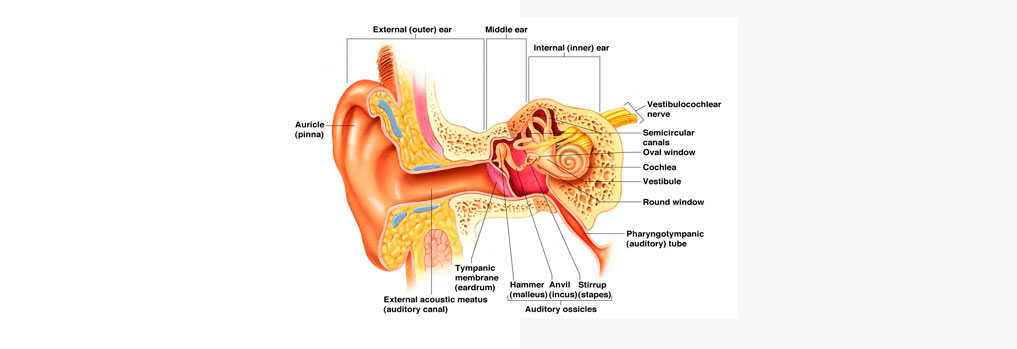
Tympanoplasty is usually performed with the patient under general anesthesia. Then the surgeon will place an instrument called an Ear speculum in the external Ear canal. The operating microscope is then positioned.
Micro Ear Surgery

Hearing is one of the special senses God has bestowed upon human beings. One can appreciate the value of hearing only when one ceases to hear.
The neurotologists in our Comprehensive Skull Base Program are specialists in medical and surgical care for disorders of the Ear, Cranial Nerves, and the Skull base. They treat patients of all ages for hearing loss, balance problems, tumors, infections, injuries, congenital (present since birth) defects, and facial nerve disorders.
Defects in the tympanic membrane or disease in mastoid bone lead to ear discharge and deafness. This mastoid bone disease triggers the risk of spreading infection to the brain and other vital nerves. One could avoid this danger with Micro Ear Surgery. It is performed with the aid of German Microscope OPMI 111 and Medtronic Visao Disposable drills. It causes near zero complications and has 99.9% success rate. The additional use of Robotic laser helps in faster recovery.
What is Microscopic Ear Surgery?
Oftentimes, perforations in the Ear Drum occur in Children. This may happen due to infection or as a result of injury. This is also called tympanic membrane perforation. Depending on your condition, your doctor may recommend Microscopic Ear Surgery. One such Surgery is called- Tympanoplasty.
Tympanoplasty is the surgical operation that is performed to reconstruct the eardrum or the small bones of the middle ear. The goal of this surgical procedure is to close the perforation and also to improve hearing.
What are the common symptoms of a Perforated Ear?
- Ear Pain
- Buzzing in the ear
- Whistling sounds when sneezing or blowing your nose
- Decrease in Hearing or Hearing Loss
- Middle Ear Inflammation or infection
When is Tympanoplasty needed?
Your Doctor may recommend Tympanoplasty is in case of
- A clear, pus-filled, or bloody Ear Drainage
- Hearing Loss
- Ringing in the Ear
- A spinning sensation from Vertigo
- Nausea or vomiting resulting from vertigo
How is the Diagnosis Done?
Diagnosis can be done in the following ways:
- Evaluating the history of the hearing loss, also for vertigo or any other facial weakness.
- Otoscopy: is usually done to inspect the mobility of the tympanic membrane and the malleus or the hammer-shaped small bone in the middle ear.
- Fistula Test: This is usually performed if the patient has a history of dizziness or the presence of a marginal perforation of the eardrum.
- Routine blood tests
- Routine urine examination
How is Tympanoplasty Performed?
The surgeon will then make an incision into the ear canal, usually made behind the ear for large perforations. The ear is then moved forward, carefully exposing the eardrum. The surgeon will lift the eardrum so that the middle ear can be examined.
If there is a hole in the eardrum, it is debrided and the abnormal area can be cut away. A piece of fascia, which is the tissue present under the skin, from the temporalis muscle, found behind the ear, is then cut and placed under the hole in the eardrum to create a new intact eardrum. This tissue, which is called a graft, allows the normal eardrum skin to grow across the hole. Sometimes, the surgeon might perform the reconstruction of the middle ear bones at this time.
The patient can return home within a couple of hours. The doctors might prescribe antibiotics and a mild pain reliever.
How Long Does the Tympanoplasty Procedure Take?
The procedure takes anywhere from 30 minutes to an hour to complete. More serious cases could take longer.
What’s the follow-up?
After 10 days, the packing is removed and the ear is checked to see if the graft was successful. As the graft must be free of infection to heal completely, antibiotics might be prescribed. If there are allergies or a cold, antibiotics and a decongestant are also prescribed.
After a month, all the packing is completely removed under the operating microscope. It is then determined whether the graft has been fully taken.
The doctor might suggest the following preventive measures to minimize the pressure that can potentially dislodge the graft.
- Avoid water from the ear
- Avoid nose blowing
- Avoid using a straw to drink
- Avoid sneezing with a shut mouth
A complete hearing test is performed four to six weeks after the surgery.
What are the risks?
There might be a few risk factors in rare cases.
- Excessive bleeding
- Infection
- Bleeding
- Reactions to medicines
- Dizziness or vertigo
- Worsening of hearing or a complete loss of hearing

